

Which Of The Following Statements Concerning The Process Of Tissue Repair Is Falss
Chapter 14. The Creature Torso: Basic Class and Office
14.ii Creature Main Tissues
Learning Objectives
By the end of this section, y'all volition be able to:
- Describe epithelial tissues
- Discuss the different types of connective tissues in animals
- Describe three types of muscle tissues
- Describe nervous tissue
Multicellular, complex animals accept four main types of tissue: epithelial, connective, muscle, and nervous. Recall that tissues are groups of similar cells carrying out related functions. These tissues combine to form organs—like the skin or kidney—that have specific, specialized functions within the trunk. Organs are organized into organ systems to perform functions; examples include the circulatory system, which consists of the heart and blood vessels, and the digestive organization, consisting of several organs, including the breadbasket, intestines, liver, and pancreas. Organ systems come up together to create an unabridged organism.
Epithelial Tissues
Epithelial tissues cover the outside of organs and structures in the trunk and line the lumens of organs in a unmarried layer or multiple layers of cells. The types of epithelia are classified past the shapes of cells present and the number of layers of cells. Epithelia composed of a single layer of cells are called simple epithelia; epithelial tissue composed of multiple layers is called stratified epithelia. Table 14.two summarizes the different types of epithelial tissues.
| Jail cell shape | Description | Location |
|---|---|---|
| squamous | flat, irregular round shape | simple: lung alveoli, capillaries stratified: skin, oral fissure, vagina |
| cuboidal | cube shaped, fundamental nucleus | glands, renal tubules |
| columnar | tall, narrow, nucleus toward base tall, narrow, nucleus forth cell | elementary: digestive tract pseudostratified: respiratory tract |
| transitional | round, simple but appear stratified | urinary float |
Squamous Epithelia
Squamous epithelial cells are by and large round, apartment, and accept a modest, centrally located nucleus. The cell outline is slightly irregular, and cells fit together to form a roofing or lining. When the cells are arranged in a single layer (simple epithelia), they facilitate diffusion in tissues, such every bit the areas of gas exchange in the lungs and the exchange of nutrients and waste at blood capillaries.
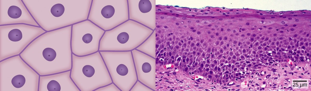
Effigy fourteen.vii a illustrates a layer of squamous cells with their membranes joined together to form an epithelium. Epitome Figure 14.vii b illustrates squamous epithelial cells bundled in stratified layers, where protection is needed on the torso from exterior abrasion and damage. This is called a stratified squamous epithelium and occurs in the skin and in tissues lining the mouth and vagina.
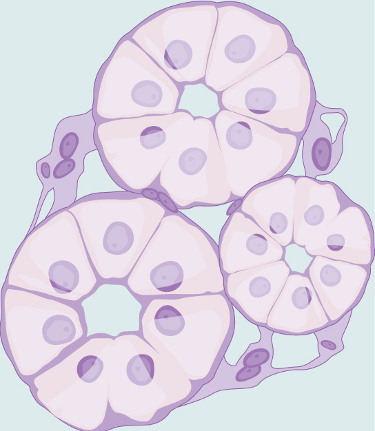
Cuboidal Epithelia
Cuboidal epithelial cells, shown in Figure 14.8, are cube-shaped with a single, central nucleus. They are most commonly found in a single layer representing a elementary epithelia in glandular tissues throughout the body where they prepare and secrete glandular material. They are also constitute in the walls of tubules and in the ducts of the kidney and liver.
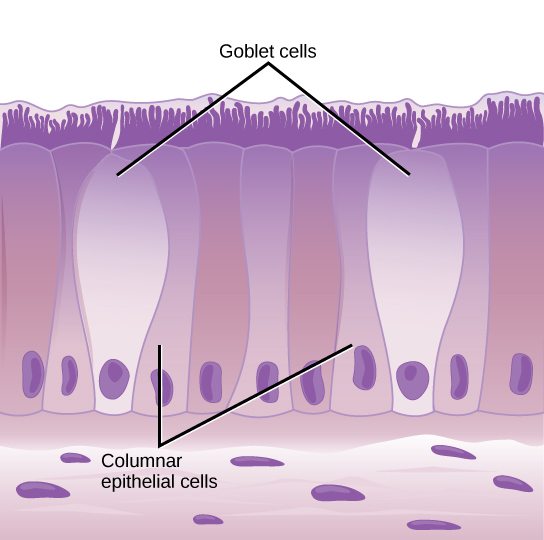
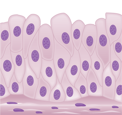
Columnar Epithelia
Columnar epithelial cells are taller than they are wide: they resemble a stack of columns in an epithelial layer, and are most ordinarily plant in a single-layer arrangement. The nuclei of columnar epithelial cells in the digestive tract appear to be lined upwardly at the base of the cells, every bit illustrated in Figure 14.9. These cells absorb material from the lumen of the digestive tract and gear up information technology for entry into the body through the circulatory and lymphatic systems.
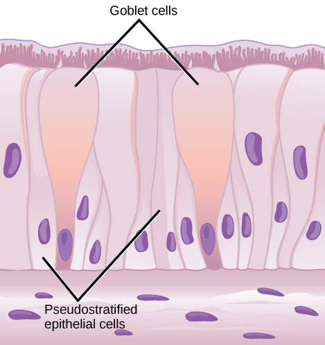
Columnar epithelial cells lining the respiratory tract appear to exist stratified. Yet, each cell is attached to the base of operations membrane of the tissue and, therefore, they are simple tissues. The nuclei are arranged at unlike levels in the layer of cells, making information technology appear every bit though there is more than i layer, every bit seen in Figure fourteen.10. This is chosen pseudostratified, columnar epithelia. This cellular covering has cilia at the apical, or free, surface of the cells. The cilia heighten the motion of mucous and trapped particles out of the respiratory tract, helping to protect the organisation from invasive microorganisms and harmful cloth that has been breathed into the body. Goblet cells are interspersed in some tissues (such as the lining of the trachea). The goblet cells incorporate mucous that traps irritants, which in the example of the trachea proceed these irritants from getting into the lungs.
Transitional Epithelia
Transitional or uroepithelial cells appear just in the urinary system, primarily in the bladder and ureter. These cells are arranged in a stratified layer, only they have the capability of appearing to pile upward on peak of each other in a relaxed, empty float, as illustrated in Figure xiv.11. Every bit the urinary bladder fills, the epithelial layer unfolds and expands to hold the book of urine introduced into information technology. As the bladder fills, it expands and the lining becomes thinner. In other words, the tissue transitions from thick to thin.
Which of the following statements about types of epithelial cells is fake?
- Simple columnar epithelial cells line the tissue of the lung.
- Elementary cuboidal epithelial cells are involved in the filtering of blood in the kidney.
- Pseudostratisfied columnar epithilia occur in a single layer, but the arrangement of nuclei makes information technology appear that more than one layer is present.
- Transitional epithelia alter in thickness depending on how full the bladder is.
Connective Tissues
Connective tissues are made up of a matrix consisting of living cells and a non-living substance, chosen the footing substance. The basis substance is made of an organic substance (usually a poly peptide) and an inorganic substance (usually a mineral or water). The principal cell of connective tissues is the fibroblast. This cell makes the fibers establish in virtually all of the connective tissues. Fibroblasts are motile, able to conduct out mitosis, and can synthesize whichever connective tissue is needed. Macrophages, lymphocytes, and, occasionally, leukocytes tin exist found in some of the tissues. Some tissues have specialized cells that are non found in the others. The matrix in connective tissues gives the tissue its density. When a connective tissue has a loftier concentration of cells or fibers, it has proportionally a less dumbo matrix.
The organic portion or protein fibers found in connective tissues are either collagen, elastic, or reticular fibers. Collagen fibers provide strength to the tissue, preventing information technology from being torn or separated from the surrounding tissues. Elastic fibers are made of the protein elastin; this fiber tin can stretch to one and 1 half of its length and return to its original size and shape. Elastic fibers provide flexibility to the tissues. Reticular fibers are the tertiary type of protein fiber found in connective tissues. This cobweb consists of sparse strands of collagen that form a network of fibers to back up the tissue and other organs to which it is connected. The various types of connective tissues, the types of cells and fibers they are made of, and sample locations of the tissues is summarized in Table fourteen.3.
| Tissue | Cells | Fibers | Location |
|---|---|---|---|
| loose/areolar | fibroblasts, macrophages, some lymphocytes, some neutrophils | few: collagen, rubberband, reticular | around blood vessels; anchors epithelia |
| dumbo, fibrous connective tissue | fibroblasts, macrophages, | mostly collagen | irregular: pare regular: tendons, ligaments |
| cartilage | chondrocytes, chondroblasts | hyaline: few collagen fibrocartilage: large amount of collagen | shark skeleton, fetal bones, homo ears, intervertebral discs |
| bone | osteoblasts, osteocytes, osteoclasts | some: collagen, elastic | vertebrate skeletons |
| adipose | adipocytes | few | adipose (fat) |
| blood | red claret cells, white claret cells | none | blood |
Loose/Areolar Connective Tissue
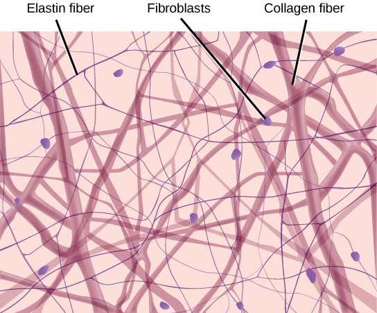
Loose connective tissue, also called areolar connective tissue, has a sampling of all of the components of a connective tissue. As illustrated in Figure fourteen.12, loose connective tissue has some fibroblasts; macrophages are nowadays also. Collagen fibers are relatively wide and stain a lite pink, while rubberband fibers are thin and stain night blue to blackness. The infinite between the formed elements of the tissue is filled with the matrix. The material in the connective tissue gives information technology a loose consistency like to a cotton ball that has been pulled apart. Loose connective tissue is establish effectually every blood vessel and helps to keep the vessel in identify. The tissue is also institute effectually and betwixt most body organs. In summary, areolar tissue is tough, yet flexible, and comprises membranes.
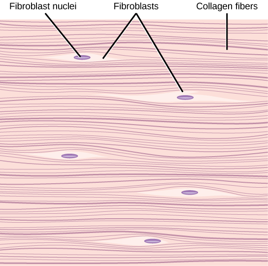
Fibrous Connective Tissue
Fibrous connective tissues contain big amounts of collagen fibers and few cells or matrix textile. The fibers can be bundled irregularly or regularly with the strands lined up in parallel. Irregularly arranged fibrous connective tissues are plant in areas of the body where stress occurs from all directions, such as the dermis of the skin. Regular fibrous connective tissue, shown in Figure 14.13, is plant in tendons (which connect muscles to bones) and ligaments (which connect bones to bones).
Cartilage
Cartilage is a connective tissue with a big amount of the matrix and variable amounts of fibers. The cells, chosen chondrocytes, make the matrix and fibers of the tissue. Chondrocytes are constitute in spaces inside the tissue chosen lacunae.
A cartilage with few collagen and elastic fibers is hyaline cartilage, illustrated in Figure fourteen.14. The lacunae are randomly scattered throughout the tissue and the matrix takes on a milky or scrubbed appearance with routine histological stains. Sharks have cartilaginous skeletons, every bit does about the entire human skeleton during a specific pre-nascency developmental stage. A remnant of this cartilage persists in the outer portion of the human nose. Hyaline cartilage is likewise institute at the ends of long bones, reducing friction and cushioning the articulations of these bones.
Rubberband cartilage has a large amount of rubberband fibers, giving it tremendous flexibility. The ears of near vertebrate animals contain this cartilage as do portions of the larynx, or voice box. Fibrocartilage contains a large amount of collagen fibers, giving the tissue tremendous forcefulness. Fibrocartilage comprises the intervertebral discs in vertebrate animals. Hyaline cartilage plant in movable joints such as the knee and shoulder becomes damaged equally a outcome of age or trauma. Damaged hyaline cartilage is replaced by fibrocartilage and results in the joints condign "potent."
Bone
Bone, or osseous tissue, is a connective tissue that has a large amount of two different types of matrix cloth. The organic matrix is similar to the matrix material found in other connective tissues, including some amount of collagen and elastic fibers. This gives strength and flexibility to the tissue. The inorganic matrix consists of mineral salts—mostly calcium salts—that give the tissue hardness. Without acceptable organic material in the matrix, the tissue breaks; without adequate inorganic material in the matrix, the tissue bends.
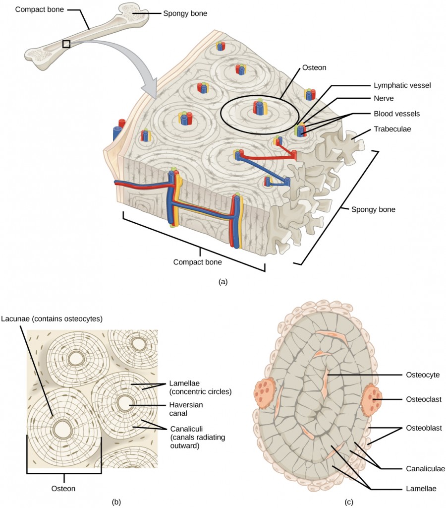
There are three types of cells in bone: osteoblasts, osteocytes, and osteoclasts. Osteoblasts are active in making os for growth and remodeling. Osteoblasts deposit bone material into the matrix and, after the matrix surrounds them, they continue to live, simply in a reduced metabolic state every bit osteocytes. Osteocytes are found in lacunae of the os. Osteoclasts are active in breaking downwards bone for os remodeling, and they provide access to calcium stored in tissues. Osteoclasts are usually plant on the surface of the tissue.
Bone can exist divided into two types: compact and spongy. Compact bone is found in the shaft (or diaphysis) of a long bone and the surface of the apartment basic, while spongy bone is found in the terminate (or epiphysis) of a long bone. Compact bone is organized into subunits chosen osteons, as illustrated in Figure 14.15. A claret vessel and a nervus are establish in the center of the construction within the Haversian culvert, with radiating circles of lacunae around it known as lamellae. The wavy lines seen betwixt the lacunae are microchannels chosen canaliculi; they connect the lacunae to aid diffusion between the cells. Spongy bone is made of tiny plates called trabeculae these plates serve as struts to requite the spongy os strength. Over time, these plates can break causing the bone to become less resilient. Bone tissue forms the internal skeleton of vertebrate animals, providing structure to the animal and points of attachment for tendons.
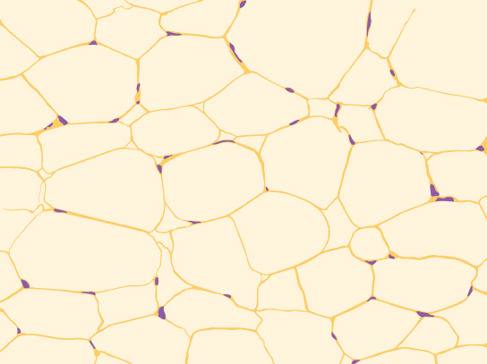
Adipose Tissue
Adipose tissue, or fatty tissue, is considered a connective tissue even though information technology does not have fibroblasts or a real matrix and but has a few fibers. Adipose tissue is made up of cells called adipocytes that collect and store fat in the form of triglycerides, for energy metabolism. Adipose tissues additionally serve every bit insulation to help maintain body temperatures, allowing animals to exist endothermic, and they office every bit cushioning against impairment to body organs. Under a microscope, adipose tissue cells appear empty due to the extraction of fatty during the processing of the material for viewing, every bit seen in Figure 14.16. The thin lines in the paradigm are the prison cell membranes, and the nuclei are the small, black dots at the edges of the cells.
Blood
Claret is considered a connective tissue considering information technology has a matrix, as shown in Figure xiv.17. The living prison cell types are red blood cells (RBC), too called erythrocytes, and white blood cells (WBC), as well chosen leukocytes. The fluid portion of whole blood, its matrix, is commonly called plasma.
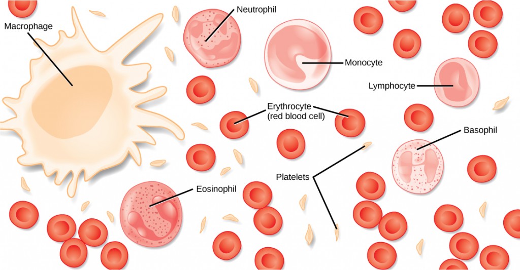
The prison cell found in greatest abundance in blood is the erythrocyte. Erythrocytes are counted in millions in a claret sample: the average number of cherry-red blood cells in primates is 4.7 to five.5 million cells per microliter. Erythrocytes are consistently the aforementioned size in a species, but vary in size between species. For example, the average diameter of a primate red blood cell is 7.5 µl, a dog is close at seven.0 µl, merely a cat's RBC diameter is five.9 µl. Sheep erythrocytes are fifty-fifty smaller at 4.six µl. Mammalian erythrocytes lose their nuclei and mitochondria when they are released from the bone marrow where they are fabricated. Fish, amphibian, and avian red blood cells maintain their nuclei and mitochondria throughout the cell's life. The main job of an erythrocyte is to carry and evangelize oxygen to the tissues.
Leukocytes are the predominant white claret cells constitute in the peripheral blood. Leukocytes are counted in the thousands in the blood with measurements expressed every bit ranges: primate counts range from 4,800 to x,800 cells per µl, dogs from 5,600 to xix,200 cells per µl, cats from eight,000 to 25,000 cells per µl, cattle from 4,000 to 12,000 cells per µl, and pigs from 11,000 to 22,000 cells per µl.
Lymphocytes function primarily in the allowed response to foreign antigens or material. Different types of lymphocytes make antibodies tailored to the foreign antigens and control the production of those antibodies. Neutrophils are phagocytic cells and they participate in 1 of the early on lines of defence force against microbial invaders, aiding in the removal of bacteria that has entered the body. Another leukocyte that is establish in the peripheral blood is the monocyte. Monocytes give ascent to phagocytic macrophages that clean upwards expressionless and damaged cells in the body, whether they are foreign or from the host animal. Two additional leukocytes in the claret are eosinophils and basophils—both help to facilitate the inflammatory response.
The slightly granular material among the cells is a cytoplasmic fragment of a cell in the bone marrow. This is called a platelet or thrombocyte. Platelets participate in the stages leading up to coagulation of the blood to stop bleeding through damaged blood vessels. Blood has a number of functions, but primarily it transports textile through the body to bring nutrients to cells and remove waste product material from them.
Muscle Tissues
There are three types of muscle in brute bodies: smooth, skeletal, and cardiac. They differ by the presence or absence of striations or bands, the number and location of nuclei, whether they are voluntarily or involuntarily controlled, and their location within the body. Table 14.4 summarizes these differences.
| Type of Muscle | Striations | Nuclei | Control | Location |
|---|---|---|---|---|
| shine | no | single, in center | involuntary | visceral organs |
| skeletal | yes | many, at periphery | voluntary | skeletal muscles |
| cardiac | yes | unmarried, in center | involuntary | center |
Smoothen Musculus
Smooth muscle does non have striations in its cells. It has a single, centrally located nucleus, as shown in Figure 14.18. Constriction of smooth muscle occurs under involuntary, autonomic nervous control and in response to local conditions in the tissues. Smoothen muscle tissue is also called non-striated as it lacks the banded advent of skeletal and cardiac musculus. The walls of blood vessels, the tubes of the digestive system, and the tubes of the reproductive systems are equanimous of mostly smooth musculus.
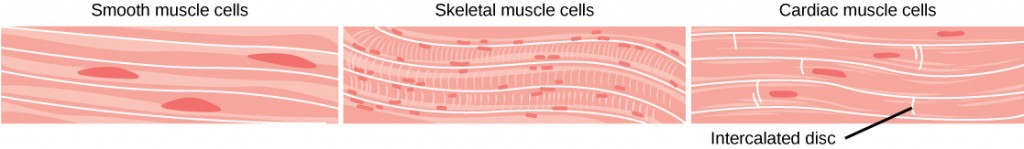
Skeletal Muscle
Skeletal muscle has striations across its cells acquired by the system of the contractile proteins actin and myosin. These musculus cells are relatively long and have multiple nuclei along the border of the prison cell. Skeletal musculus is under voluntary, somatic nervous system command and is constitute in the muscles that motility bones. Figure 14.eighteen illustrates the histology of skeletal muscle.
Cardiac Musculus
Cardiac muscle, shown in Effigy 14.18, is found only in the heart. Similar skeletal musculus, it has cross striations in its cells, but cardiac muscle has a unmarried, centrally located nucleus. Cardiac muscle is non under voluntary control but tin be influenced past the autonomic nervous system to speed up or slow downwardly. An added characteristic to cardiac muscle cells is a line than extends along the terminate of the jail cell as it abuts the next cardiac cell in the row. This line is called an intercalated disc: it assists in passing electrical impulse efficiently from 1 cell to the adjacent and maintains the stiff connectedness betwixt neighboring cardiac cells.
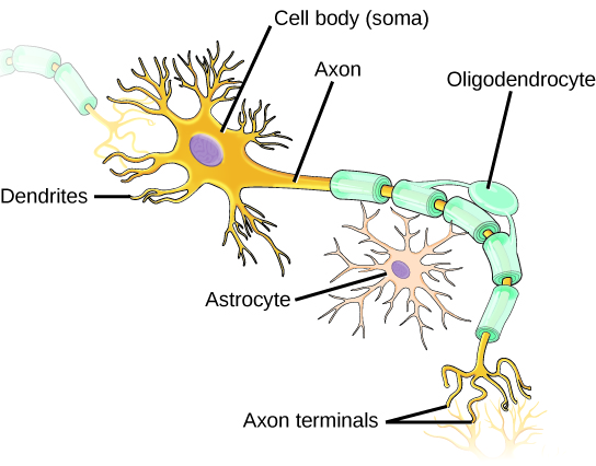
Nervous Tissues
Nervous tissues are fabricated of cells specialized to receive and transmit electric impulses from specific areas of the body and to transport them to specific locations in the torso. The master cell of the nervous system is the neuron, illustrated in Figure 14.nineteen. The large structure with a central nucleus is the cell torso of the neuron. Projections from the jail cell body are either dendrites specialized in receiving input or a single axon specialized in transmitting impulses. Some glial cells are besides shown. Astrocytes regulate the chemical environment of the nerve jail cell, and oligodendrocytes insulate the axon and then the electrical nerve impulse is transferred more than efficiently. Other glial cells that are not shown back up the nutritional and waste matter requirements of the neuron. Some of the glial cells are phagocytic and remove debris or damaged cells from the tissue. A nervus consists of neurons and glial cells.
Concept in Activeness

Click through the interactive review to learn more than nigh epithelial tissues.
Pathologist
A pathologist is a medical dr. or veterinary who has specialized in the laboratory detection of affliction in animals, including humans. These professionals consummate medical school education and follow information technology with an extensive post-graduate residency at a medical centre. A pathologist may oversee clinical laboratories for the evaluation of trunk tissue and blood samples for the detection of illness or infection. They examine tissue specimens through a microscope to identify cancers and other diseases. Some pathologists perform autopsies to determine the cause of death and the progression of disease.
Exercises
- Which of the following statements almost types of epithelial cells is false?
- Simple columnar epithelial cells line the tissue of the lung.
- Simple cuboidal epithelial cells are involved in the filtering of blood in the kidney.
- Pseudostratisfied columnar epithilia occur in a single layer, but the arrangement of nuclei makes it announced that more than one layer is present.
- Transitional epithelia change in thickness depending on how full the bladder is.
- State whether each of the post-obit processes are regulated by a positive feedback loop or a negative feedback loop.
- A person feels satiated after eating a large meal.
- The claret has plenty of cherry blood cells. As a result, erythropoietin, a hormone that stimulates the product of new red blood cells, is no longer released from the kidney.
- When bacteria are destroyed past leuckocytes, pyrogens are released into the blood. Pyrogens reset the trunk's thermostat to a higher temperature, resulting in fever. How might pyrogens cause the trunk temperature to ascension?
- Which blazon of creature maintains a constant internal trunk temperature?
- endotherm
- ectotherm
- coelomate
- mesoderm
- The symmetry constitute in animals that move swiftly is ________.
- radial
- bilateral
- sequential
- interrupted
- What term describes the status of a desert mouse that lowers its metabolic rate and "sleeps" during the hot day?
- turgid
- hibernation
- estivation
- normal sleep design
- A plane that divides an beast into equal right and left portions is ________.
- diagonal
- midsagittal
- coronal
- transverse
- A plane that divides an animal into dorsal and ventral portions is ________.
- sagittal
- midsagittal
- coronal
- transverse
- The pleural cavity is a part of which cavity?
- dorsal cavity
- thoracic crenel
- abdominal cavity
- pericardial crenel
- Which type of epithelial cell is all-time adjusted to aid diffusion?
- squamous
- cuboidal
- columnar
- transitional
- Which type of epithelial jail cell is constitute in glands?
- squamous
- cuboidal
- columnar
- transitional
- Which type of epithelial cell is found in the urinary bladder?
- squamous
- cuboidal
- columnar
- transitional
- Which type of connective tissue has the most fibers?
- loose connective tissue
- fibrous connective tissue
- cartilage
- bone
- Which type of connective tissue has a mineralized different matrix?
- loose connective tissue
- fibrous connective tissue
- cartilage
- bone
- The cell found in bone that breaks it down is chosen an ________.
- osteoblast
- osteocyte
- osteoclast
- osteon
- The cell plant in bone that makes the bone is chosen an ________.
- osteoblast
- osteocyte
- osteoclast
- osteon
- Plasma is the ________.
- fibers in blood
- matrix of claret
- cell that phagocytizes leaner
- jail cell fragment found in the tissue
- The blazon of muscle cell nether voluntary control is the ________.
- smooth muscle
- skeletal muscle
- cardiac muscle
- visceral muscle
- The part of a neuron that contains the nucleus is the
- cell body
- dendrite
- axon
- glial
- When faced with a sudden driblet in environmental temperature, an endothermic animal will:
- feel a drop in its torso temperature
- wait to meet if information technology goes lower
- increment musculus activity to generate heat
- add together fur or fat to increment insulation
- Which is an example of negative feedback?
- lowering of blood glucose subsequently a meal
- blood clotting afterwards an injury
- lactation during nursing
- uterine contractions during labor
- Which method of heat commutation occurs during direct contact between the source and animal?
- radiation
- evaporation
- convection
- conduction
- The body's thermostat is located in the ________.
- homeostatic receptor
- hypothalamus
- medulla
- vasodilation center
- How does diffusion limit the size of an organism? How is this counteracted?
- What is the relationship between BMR and body size? Why?
- How tin can squamous epithelia both facilitate diffusion and forbid damage from chafe?
- What are the similarities between cartilage and bone?
- Why are negative feedback loops used to control body homeostasis?
- Why is a fever a "good thing" during a bacterial infection?
- How is a condition such every bit diabetes a good example of the failure of a ready point in humans?
Answers
- A
- Both processes are the result of negative feedback loops. Negative feedback loops, which tend to go on a arrangement at equilibrium, are more mutual than positive feedback loops.
- Pyrogens increase body temperature by causing the blood vessels to constrict, inducing shivering, and stopping sweat glands from secreting fluid.
- A
- B
- C
- B
- D
- B
- C
- B
- D
- B
- D
- C
- A
- B
- B
- B
- C
- A
- D
- B
- Diffusion is effective over a very curt distance. If a cell exceeds this distance in its size, the heart of the cell cannot get adequate nutrients nor can it expel plenty waste to survive. To compensate for this, cells can loosely adhere to each other in a liquid medium, or develop into multi-celled organisms that use circulatory and respiratory systems to deliver nutrients and remove wastes.
- Basal Metabolic Rate is an expression of the metabolic processes that occur to maintain an private's functioning and body temperature. Smaller bodied animals have a relatively big surface area compared to a much larger animal. The large beast's large surface surface area leads to increased oestrus loss that the beast must compensate for, resulting in a college BMR. A small animal, having less relative surface area, does non lose as much heat and has a correspondingly lower BMR.
- Squamous epithelia can exist either simple or stratified. As a single layer of cells, information technology presents a very thin epithelia that minimally inhibits diffusion. As a stratified epithelia, the surface cells tin can be sloughed off and the cells in deeper layers protect the underlying tissues from damage.
- Both contain cells other than the traditional fibroblast. Both take cells that lodge in spaces within the tissue called lacunae. Both collagen and elastic fibers are constitute in bone and cartilage. Both tissues participate in vertebrate skeletal evolution and formation.
- An adjustment to a change in the internal or external surroundings requires a alter in the management of the stimulus. A negative feedback loop accomplishes this, while a positive feedback loop would continue the stimulus and result in damage to the beast.
- Mammalian enzymes increase action to the bespeak of denaturation, increasing the chemic activity of the cells involved. Bacterial enzymes have a specific temperature for their most efficient activity and are inhibited at either higher or lower temperatures. Fever results in an increase in the destruction of the invading bacteria past increasing the effectiveness of body defenses and an inhibiting bacterial metabolism.
- Diabetes is often associated with a lack in product of insulin. Without insulin, blood glucose levels go up subsequently a repast, just never get back downwardly to normal levels.
Glossary
cartilage: type of connective tissue with a big corporeality of footing substance matrix, cells called chondrocytes, and some amount of fibers
chondrocyte: cell plant in cartilage
columnar epithelia: epithelia made of cells taller than they are wide, specialized in assimilation
connective tissue: type of tissue made of cells, ground substance matrix, and fibers
cuboidal epithelia: epithelia made of cube-shaped cells, specialized in glandular functions
dorsal cavity: trunk crenel on the posterior or back portion of an animal; includes the cranial and vertebral cavities
ectotherm: animal incapable of maintaining a relatively constant internal body temperature
endotherm: brute capable of maintaining a relatively constant internal body temperature
epithelial tissue: tissue that either lines or covers organs or other tissues
estivation: torpor in response to extremely loftier temperatures and low water availability
fibrous connective tissue: type of connective tissue with a high concentration of fibers
hibernation: torpor over a long menstruation of time, such as a wintertime
homeostasis: dynamic equilibrium maintaining appropriate body functions
lacuna: space in cartilage and bone that contains living cells
matrix: component of connective tissue made of both living and non-living (ground substances) cells
negative feedback loop: feedback to a command mechanism that increases or decreases a stimulus instead of maintaining it
osteon: subunit of compact bone
positive feedback loop: feedback to a control mechanism that continues the management of a stimulus
pseudostratified: layer of epithelia that appears multilayered, but is a elementary covering
prepare point: midpoint or target point in homeostasis
simple epithelia: unmarried layer of epithelial cells
squamous epithelia: type of epithelia made of flat cells, specialized in aiding improvidence or preventing abrasion
stratified epithelia: multiple layers of epithelial cells
torpor: decrease in activeness and metabolism that allows an creature to survive adverse conditions
trabecula: tiny plate that makes up spongy bone and gives it strength

Which Of The Following Statements Concerning The Process Of Tissue Repair Is Falss,
Source: https://opentextbc.ca/biology/chapter/14-2-animal-primary-tissues/
Posted by: raneyotion1985.blogspot.com
0 Response to "Which Of The Following Statements Concerning The Process Of Tissue Repair Is Falss"
Post a Comment